

Services
| Tooth extraction | |
| Complicated tooth extraction | |
| Surgical tooth extraction | |
| Extraction of a milk tooth | |
| Non-surgical removal of wisdom teeth | |
| Composite filling | |
| Amalgam filling | |
| Composite upgrade | |
| Composite facet | |
| Tartar removal with polishing | |
| Tartar removal with sandblasting | |
| Drug application | |
| Mechanical endodontic treatment of a multi-channel tooth | |
| Mechanical revision of the channel | |
| Metal-ceramic crown | |
| Metal-free zirconium crown | |
| Thermoplast prosthesis partial | |
| Thermoplast prosthesis total | |
| Skeletonized Vizil prosthesis WHITE VIZIL | |
| Retainer film after prosthetic work | |
| Total prosthesis Acrylic | |
| Partial prosthesis Acrylic | |
| Repair of acrylic prostheses | |
| Fixed orthodontic appliance per jaw | |
| Fixed orthodontic appliance SALE UNTIL 31.01.2023 | |
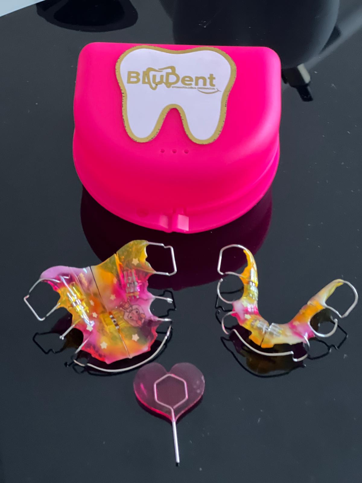
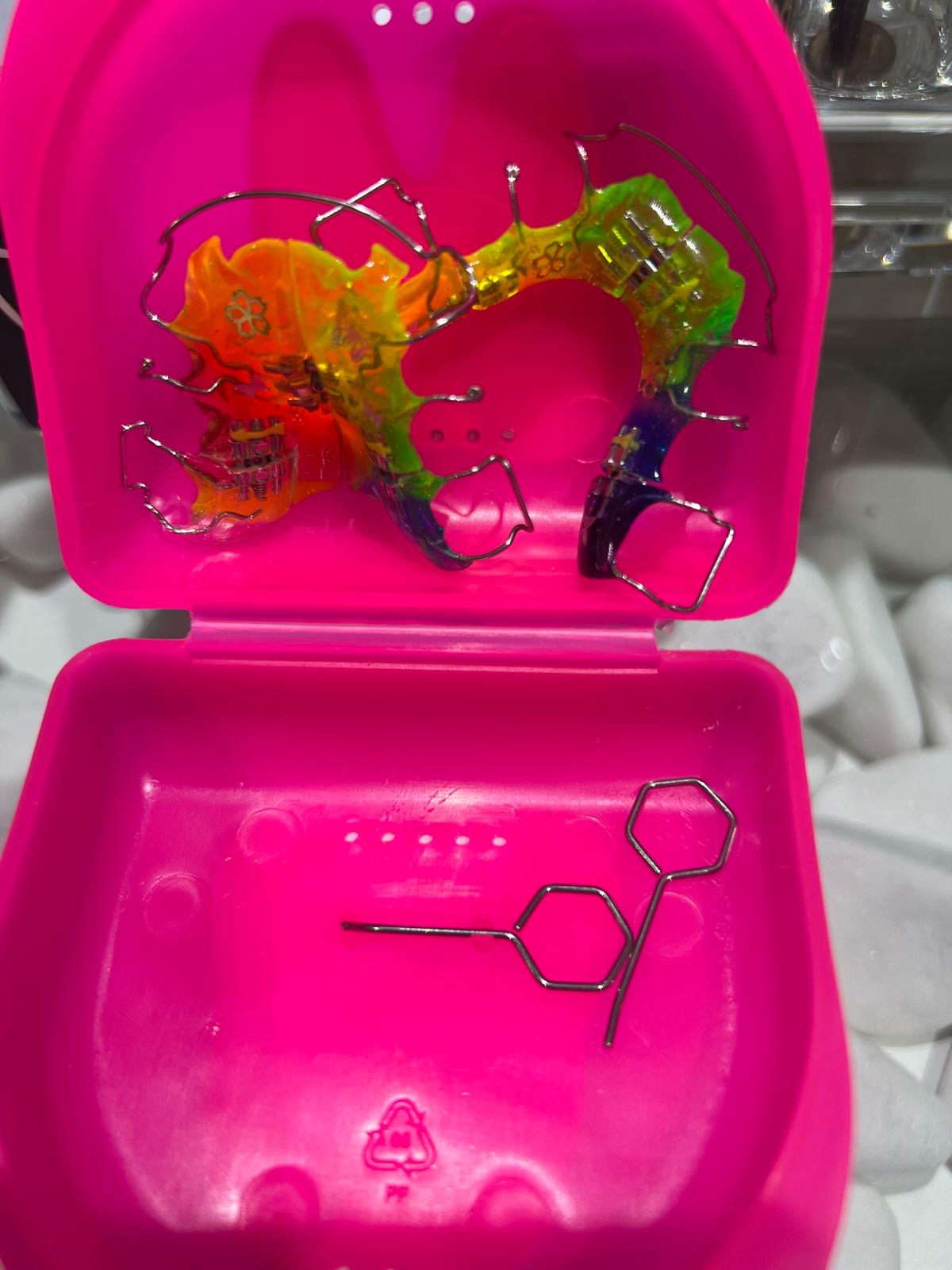
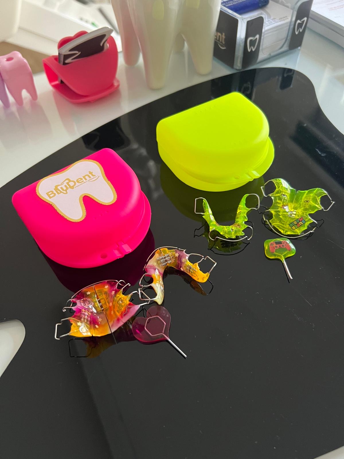
| Mobile orthodontic device | |
| Production of plinths for analysis | |
| Retainer foil | |
| Orthodontic | |
| Orthopanic recording OPT | |
| Retroalveolar imaging | |
| IMPLANT ZIMMER PREMIUM | |
| Implantation of artificial bone with membrane | |
| Gingivectomy | |
| Apicotomy | |
| Office teeth whitening | |
| Home teeth whitening with splints | |
| Teeth whitening one jaw | |
| Splint for bruxism |

ORTHOPAN
Dental X-ray – ORTHOPAN IMAGE
What is an Ortopan?
An ortopan is an X-ray image that has been completely irreplaceable in dentistry for decades. It shows the hard dental tissues and bones, as well as existing cavities (sinuses, nasal cavity). On it, the dentist can see tooth decay, bone processes, and changes in the sinuses.
3D DENTAL IMAGING
What is the difference between a regular and a 3D Ortopan image?
3D is a more modern and advanced version that allows the dentist to see all dimensions much more clearly. It has become the gold standard before implant placement because it enables visualization of bone width, height, and density, as well as the relationship to surrounding anatomical structures such as the mandibular canal, sinus floor, etc. These are all essential data for successful implant placement.
Cephalogram – TELERADIOGRAPH
Lateral cephalogram
A teleradiograph, or profile X-ray, is most often used when establishing an orthodontic diagnosis and creating a treatment plan, but also as a control image during and after orthodontic treatment. It shows the bony profile of the head and the contours of the soft tissues of the facial profile. On this image, orthodontists perform cephalometric measurements (cephalometry) and, based on them, various analyses (cephalometric analyses).


Mobile Orthodontic Appliances